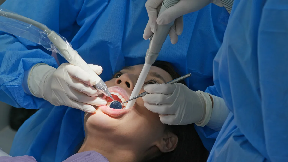










The assessment begins with diagnostic imaging. A panoramic X-ray provides an overview of all the dental and bone structures. A 3D cone beam CT scan produces a detailed three-dimensional image that helps assess bone density, root shape and root position.
Analysis of these findings guides the choice of extraction technique. The practitioner identifies potential risks and selects the appropriate instruments. A treatment plan is defined for each clinical situation.